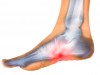
The sinus tarsi is the cavity on the lateral (outer) side of the foot in front of the ankle. The sinus tarsi space is filled with many connective tissues that contribute to the stability and the proprioception of the ankle (proprioception is the unconscious perception of movement and spatial orientation arising from stimuli within the body itself).
In sinus tarsi syndrome, the ligaments around the sinus tarsi—the interosseous and cervical ligaments—are injured, causing instability of the subtalar joint. This joint allows inversion and eversion of the foot, which is the ability to move the foot in toward the body (pronation) or out away from the body (supination).
With instability of the subtalar joint, these movements are exaggerated. This causes stress across the sinus tarsi tissues, which leads to inflammation and anterolateral ankle pain characteristic of sinus tarsi syndrome.
Injury to the ligaments around the sinus tarsi can occur in athletes such as runners and dancers whose chosen sports require a lot of jumping or sudden, quick movements and sudden stops. Sinus tarsi syndrome is thought to occur after a single traumatic event or a series of ankle sprains.
Sinus Tarsi Syndrome Symptoms To Know About
Athletes with sinus tarsi syndrome often have two symptoms:
- Deep, sharp, or pinching pain along the top and/or outer side of the foot and ankle when the foot is dorsiflexed, such as when walking up stairs; the pain may increase with time on the feet and be relieved by rest.
- A feeling of unsteadiness when walking on uneven surfaces.
Affected athletes may report having a previous ankle injury or one or more ankle sprains. An athlete with recurrent ankle sprains may have sinus tarsi syndrome.
What Causes Sinus Tarsi Syndrome?
The most common cause of sinus tarsi syndrome is trauma (in 70 percent of cases); inflammatory conditions, ganglion cysts, and foot deformities are responsible for the remaining 30 percent of cases (Radiology, 2001).
The exact reason why sinus tarsi syndrome develops is a matter of debate. One theory suggests that scar tissue, which is part of the natural healing process, causes thickening of the joint capsule. The thickened joint capsule becomes pinched between the bones in the ankle, leading to chronic inflammation.
It has also been suggested that sinus tarsi syndrome develops after ankle inversion sprains that are not treated properly. If the sensory receptors responsible for proprioception in your ankle do not heal well after a sprain, they may not regain their pre-injury ability to sense changes in ankle position.
How Is Sinus Tarsi Syndrome Diagnosed?
Your doctor will examine your foot and perform several tests to assess the stability of the subtalar joint and surrounding joints, including the talocrural joint. You will be asked to stand so that your posture can be assessed.
Some athletes with sinus tarsi syndrome may appear to have pes planus (flat feet) due to pronation, or inward leaning, of the ankle. Your doctor will manually move your foot to test passive range of motion of the ankle and subtalar joint.
This test may reveal excessive motion or looseness of the joint. A typical sign of sinus tarsi syndrome is pain in the sinus tarsi when your foot is turned in or turned out, or if you feel pain on palpation of the area.
The muscles that cross the ankle joint will also be assessed to see whether there is any loss of strength. The unaffected foot will be evaluated to compare the differences in joint mobility between both feet and to determine whether you might have instability.
Stability of the subtalar joint can be assessed in a few ways. Your doctor may hold onto your forefoot with one hand while applying an inversion and internal rotational force to your heel with the other hand.
Or you may be asked to stand on the affected foot while keeping the other foot raised and perform rotating motions of the leg and foot to reproduce symptoms.
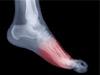
You may be referred for imaging studies. These may include radiographs of the foot, computed tomography, magnetic resonance imaging (MRI), or stress fluoroscopy—a method of visualizing the motions of the subtalar joint in real time using low-level radiation.
MRI is considered the best method to visualize the structure within the sinus tarsi, especially the interosseous and cervical ligaments. Ankle arthroscopy can also be used to evaluate the sinus tarsi for damaged tissue.
Sinus Tarsi Syndrome Treatment Options
After your doctor has evaluated you and a diagnosis of sinus tarsi syndrome has been confirmed, conservative treatment of sinus tarsi syndrome can be administered at home. Such treatment involves:
- RICE (Rest, Ice, Compression, and Elevation) to reduce inflammation and swelling in the ankle
- Identifying and eliminating the activity that may be exacerbating your subtalar joint injury to decrease the tension and thickening of the joint capsule
- Muscle-strengthening exercises to restore proper ankle proprioception
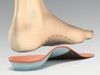
Your doctor may recommend anti-inflammatory medications, stable shoes, an ankle sleeve or brace, and over-the-counter orthotics. If your symptoms persist after conservative treatment, you may need a course of oral corticosteroids, a series of corticosteroid injections into the joint, physical therapy, or custom orthotics.
Surgery is rarely indicated, although it may be considered for patients whose symptoms do not improve after corticosteroid injections.
Most patients have two options: open surgery (through an incision for reconstruction of the subtalar joint) or closed surgery (via arthroscopic exploration).
When to Return to Play After Sinus Tarsi Syndrome
You should be able to resume your normal activities within a few days, depending on your ability to move in all directions and at appropriate speeds. You may need further treatment if symptoms return, however, in order to prevent chronic inflammation of the sinus tarsi tissues.