Over 25 percent of our bones are located in our feet. There are twenty-six bones that make up the complex structure of each foot. The foot is a strong mechanism, and it supports our entire body weight during all upright activities (standing, walking, running, etc).
It remains flexible, durable, and resilient under enormous pressure. Structurally, the foot consists of three main parts: the forefoot, midfoot, and hindfoot.
Anatomically, the bones of the foot are known as the tarsal bones, metatarsal bones, and phalanges. Let’s break down each category and discuss the importance of each bone.
Forefoot: Phalanges and Metatarsal Bones
The forefoot comprises fourteen phalanges and five metatarsal bones. One phalanx of each of the five toes connects to one of the five metatarsals.
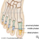
The toes are numbered one through five, starting with the big toe, which is technically known as the hallux. Some people like to call the fifth toe the “pinky.” The phalanges are referred to as follows:


- The proximal phalanx is the base of each toe and articulates the metatarsal bone and middle phalanx.
- The middle phalanx is located under the distal phalanx in the second, third, fourth and fifth toes only. The hallux does not have a middle phalanx.
- The distal phalanx is found at the end of each toe, forming the tip of the toes.
There are five metatarsals in each foot. The metatarsals are the long bones located between the tarsal bones and the phalanges.
Each metatarsal has a long shaft with two expanded ends called the base and the head. They form part of the foot’s arch and serve both as a major shock absorber and a weight-transfer mechanism.
They are connected to the forefoot and midfoot by muscles, tendons, and a special ligament known as the plantar fascia.
The metatarsals are not named individually; rather, they are numbered first through fifth, starting with the big toe. Here is a quick description of each of the metatarsal bones:
- The first metatarsal bone is the shortest and thickest.
- The second metatarsal bone is the longest and is firmly held between the first and third cuneiform bones. This bone is most susceptible to injury.
- The third metatarsal bone is in the middle and articulates the third cuneiform, as well as the second and fourth metatarsal bones.
- The fourth metatarsal bone is smaller than the third, but like the third it also articulates the surrounding metatarsals (the third and fifth), and also articulates the cuboid.
- The fifth metatarsal bone articulates the cuboid and the fourth metatarsal. Jones fractures and stress fractures are common injuries to this bone.
There are also two tiny bones located under the hallux (big toe) called sesamoid bones. Sesamoid bones develop within the tendon of a muscle to protect the tendon from wear and tear at the point where it passes over the end of the metatarsal bone.
They bear the weight of the body when the toe pushes off in walking or running. Though it is less common, sesamoid bones can be found elsewhere in the foot within other flexor tendons of the toes.
Midfoot: Lesser Tarsal Bones
The five bones that make up the midfoot are collectively known as the lesser tarsal bones. They act as an intermediary to transfer weight from the hindfoot to the forefoot with each footstep.
These bones, along with the joints and ligaments that connect them, make up most of what we would consider the arch of the foot. They include:
- The navicular bone is boat-shaped and located in the middle of the foot. It articulates with the talus and the three cuneiform bones.
- The cuboid bone is shaped like a cube and is the last of the seven tarsal bones, articulating with the fourth and fifth metatarsal bones, calcaneus, and navicular bones.
- There are three cuneiform bones in all: medial, intermediate, and lateral. They are located in the midfoot region, articulating with the corresponding metatarsal bones and navicular bone.
Hindfoot: Greater Tarsal bones
The hindfoot connects the foot to the leg at the ankle joint, and comprises two large bones called the talus and calcaneus.
These bones articulate with each other through the subtalar joint, and also link the hindfoot to the midfoot through the mid-tarsal joint. Here is a brief description of each of the tarsal bones:
- The talus connects the hindfoot to the two leg bones (the tibia and fibula) to form the ankle joint, which acts as a hinge to allow the foot to move up and down. It also links the hindfoot to the midfoot through the navicular bone to form one part of the mid-tarsal joint. From below, the talus meets up with the calcaneus to form the subtalar joint, which allows the foot to move from side to side.
- The calcaneus is also known as the heel bone, and it is the largest bone in the foot. It connects with the talus from above to form the subtalar joint. It also meets up with the cuboid bone to form the rest of the midtarsal joint. This joint allows some motion between the hindfoot and midfoot that gives the foot some flexibility